Cardiac ablation is a treatment method to correct the anomaly in the heartbeat (arrhythmia) without causing any damage to the rest of the heart. The abnormal or faulty tissue causing the problem is destroyed by exposing it to the radiofrequency energy. The treatment is given when the medicines do not turn out to be effective enough or cause side-effects.
This treatment is, more often, done using catheters (long, flexible tubes) and sometimes through open heart surgery. When done using catheters, the treatment is called catheter ablation. Here we’ll mainly focus on catheter ablation.
Types Of Cardiac Ablation

1. Atrial flutter ablation
This procedure creates scar tissue inside heart’s right upper chamber so as to block the electrical signals causing a fluttering heartbeat and restore the normal heart rhythm.
2. Pulmonary vein isolation
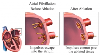
In this procedure, a scar tissue is created in heart’s left upper chamber where our four pulmonary veins (veins bringing oxygen-rich blood from lungs to heart) connect. It reduces signs and symptoms of atrial fibrillation affecting heart’s upper chambers.
3. SVT ablation
Supraventricular tachycardia or SVT ablation is a procedure that involves creation of scar tissue inside the heart. It helps in treatment of heart rhythm problems in heart’s upper chambers.
4. Ventricular tachycardia ablation
This procedure removes the heart areas in heart’s lower chambers where abnormal electrical signals crop up causing irregular or abnormally fast heartbeats.
What Happens During The Treatment?

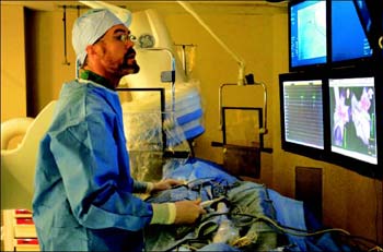
A specially trained team of doctor, nurses and technicians conduct the treatment in an electrophysiology (EP) lab or a cardiac catheterization lab.
During the procedure, small wires called electrodes are placed inside your heart to measure your heart's electrical activity.
The procedure begins by putting an IV (intravenous line) into a vein in the patient’s arm so as to get the medicine (anesthesia) to prevent pain. One can also get a medicine (sedative) to relax but will be in a conscious state. This is followed by a local anesthetic shot to numb the needle puncture site.
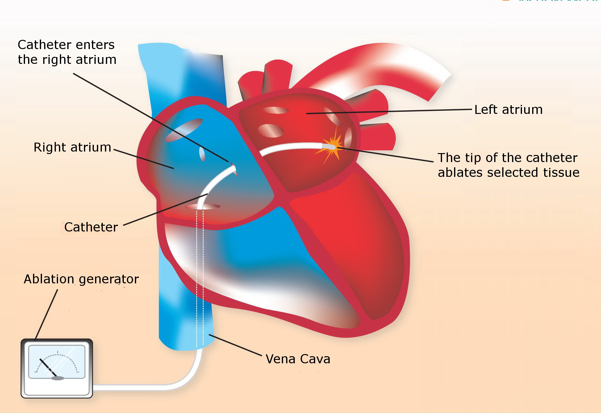
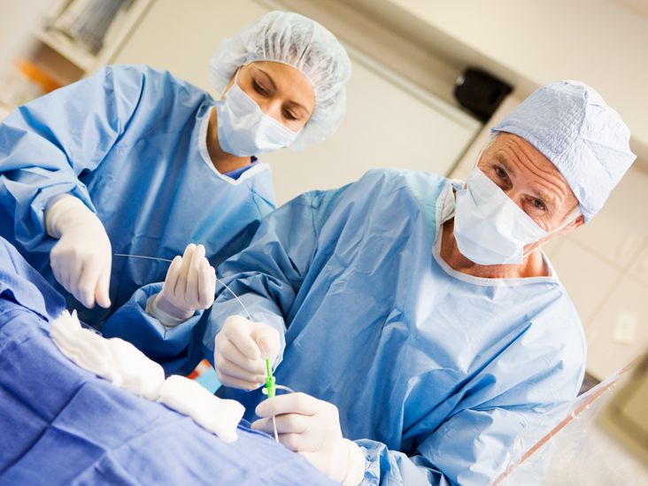
Then, the doctor makes a needle puncture through the skin and into the blood vessel (mostly veins). He guides a catheter into the blood vessel through a small tube called ‘sheath’. He uses electrode catheter to emit a small electrical pulse to look for the abnormal tissue that is causing arrhythmia.
The catheter is positioned exactly where the abnormal cells are there, inside in the heart. After that, mild and painless radiofrequency energy is sent to the tissue. As a result of this, heart muscle cells causing extra impulses and rapid heartbeats are destroyed.
The whole procedure usually takes 2-4 hours.
What Are The Risks Involved?

However, the problems that are most commonly seen problems are caused due to the catheters. Though it happens rarely, the catheter’s insertion into arteries or veins may damage the blood vessel or may lead to bleeding or infection.
How To Prepare For Catheter Ablation?

Doctors generally advise patients to stay without food and water for at least 6-8 hours before the procedure. Also, it’s best to keep the doctor informed about the medicines being taken so that he can tell whether to take them or not. In addition to that, one shouldn’t leave for home all by themselves.
What Happens After The Treatment?

The patient is moved into a recovery room. The sheath would stay on the leg for several hours after the treatment.
After the removal of the sheath, the leg needs to be kept straight for 6-8 hours. The heartbeat, pulse and blood pressure are examined. Also, Aspirin is prescribed for 2-4 weeks to reduce the risk of clotting at the ablation sites.

